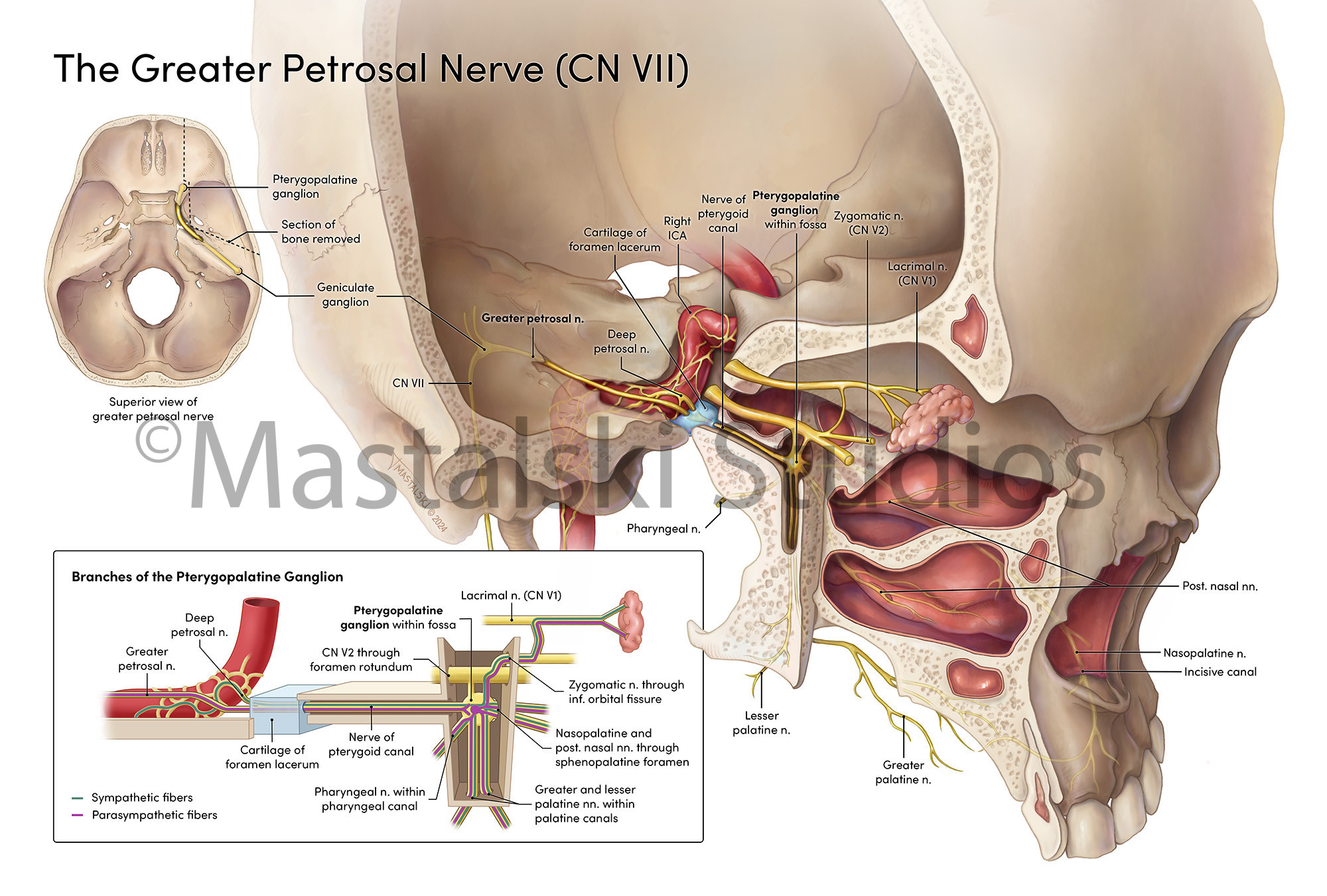
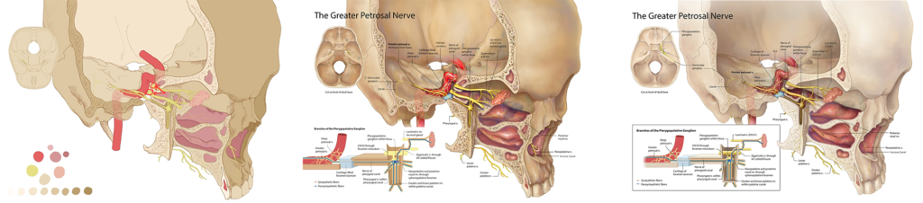
This illustration depicts the pathway of the greater petrosal nerve (GPN) within the cranium. A simplified superior view of the skull base in the upper left orients the viewer to the main image. In the main image, the pathway of the GPN can be followed from the geniculate ganglion within the petrous temporal bone to the pterygopalatine ganglion, from which fibers radiate to innervate the nasal mucosa and palate. A unique cut was planned using DICOM data to expose the fossa and path of the GPN from an oblique lateral view. A schematic inset with didactic colors illustrates the functional pathway, highlighting how parasympathetic fibers from the GPN travel alongside sympathetic fibers from the deep petrosal nerve through the pterygoid canal and branch from the pterygopalatine ganglion. For optimal contrast and legibility, all text exceeds 7:1 contrast ratio, and the color palette accommodates color blindness. Keep on scrolling to see how this piece came together!
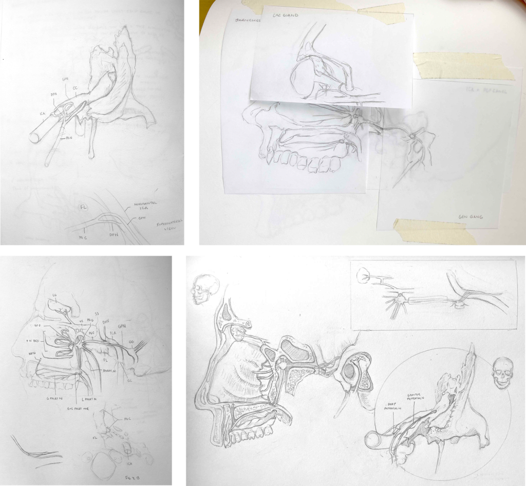
Some of my initial thumbnailing, drawn while scrolling through DICOM data, referencing skulls available in our studio, and paging through anatomical atlases
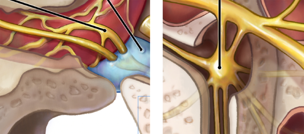
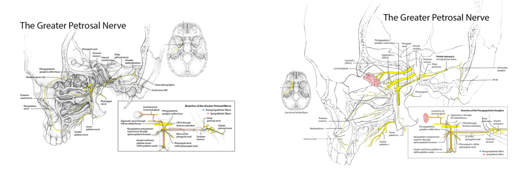
Oblique angle of skull chosen to best show everything I needed to show. At this stage, a lot of time was dedicated to using the DICOM data to lay out where I needed to cut the skull.
Playing with the composition of the three assets
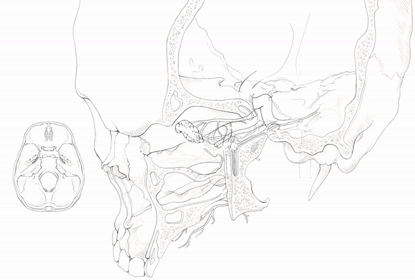
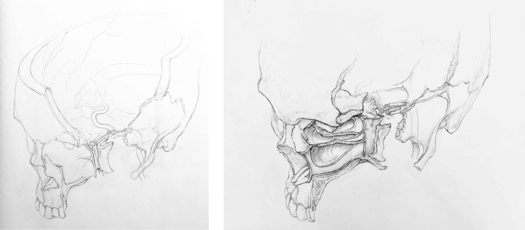
Transfer sketches of each individual asset created with graphite and compiled in Photoshop
Main image rendered in Photoshop. The skull was flipped to create a better left to right eye flow. I started with blocking in flat colors, then clipped many, many layers of shade and highlight to each block, building up the color and form over time. The schematic in the bottom left was created in Illustrator.
And here’s the final look!