Injuries to the Craniocervical Junction: A 3D Approach to Resident Education for Radiological Imaging
Preceptor: Elana Smith, MD, Associate Professor, University of Maryland School of Medicine, Diagnostic Radiology and Nuclear Medicine
Preceptor: Andrew Sinensky, MD, Resident, University of Maryland School of Medicine, Diagnostic Radiology and Nuclear Medicine
Faculty Advisor: Jeffrey Day, MD, MA, Assistant Professor, The Johns Hopkins University School of Medicine, Art as Applied to Medicine
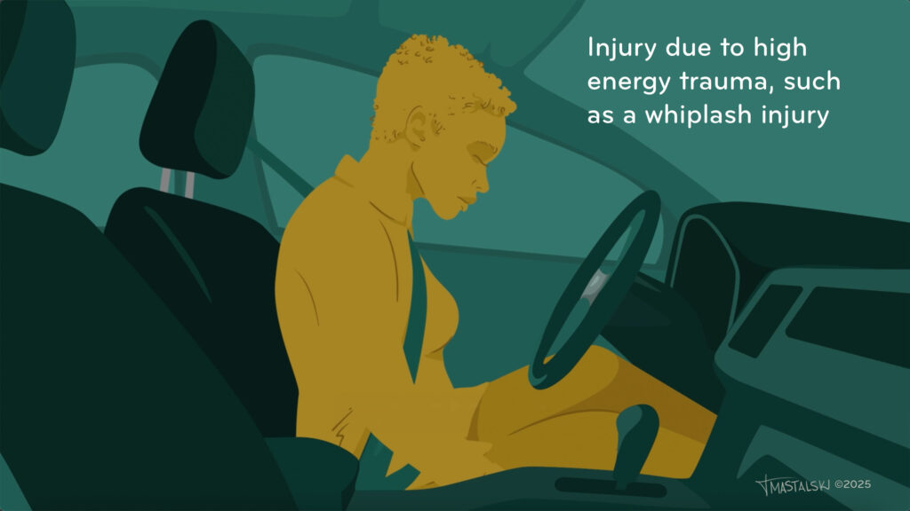
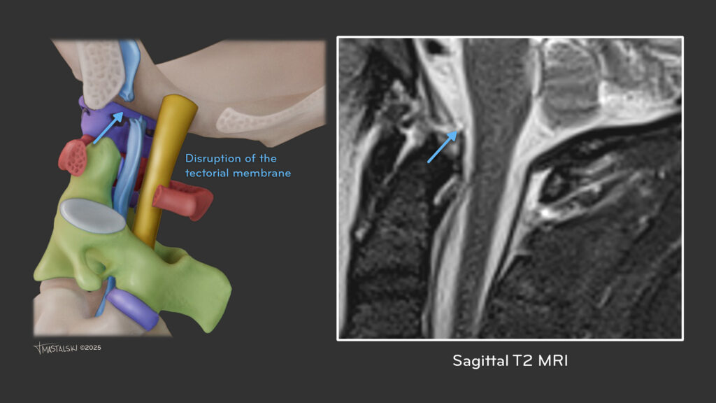
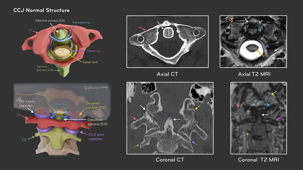
Injuries to the craniocervical junction (CCJ), due to its complex anatomy, are notoriously challenging to locate in imaging and often misdiagnosed or overlooked, leading to unnecessary extension of injuries. Difficulties in reading the imaging and a lack of resources have created a need for clear and accessible educational materials for residents. This thesis investigates the use of multimedia to assist learners in visualizing CCJ structures and injury pathologies in 3D, as well as directly relate our model to medical imaging. Testing the efficacy of our materials will provide insight into the value of multimedia for enhancing radiological education and provide new resources to facilitate efficient diagnoses of injuries.
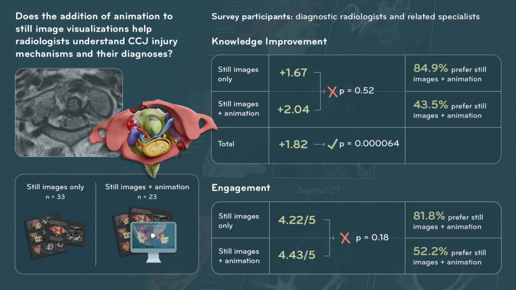
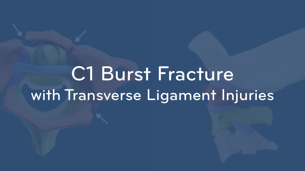
We created a series of animations highlighting CCJ injuries and still images relating them to CT/MR imaging. We tested the efficacy of the media using an online Qualtrics survey distributed to radiology-associated residencies and medical students from 10 universities and research institutions, including The Johns Hopkins University and the University of Maryland. Our main study question was if the addition of an animation to still image explanations would help learners better understand CCJ injuries and their diagnoses. Participants were randomized into two groups– still images and still images + animation. Each participant completed a pre-test, viewed a module, then completed an engagement survey, post-test, and comparison survey.
Our survey results showed significant improvement in testing among both groups (p = 0.000064), suggesting the overall efficacy of our materials. There was neither a significant difference in test scores between the still images and still images + animation groups (p = 0.52), nor a significant difference in total engagement survey scores (p = 0.18) with the still images + animation group rating higher engagement. However, participants from both groups overwhelmingly chose still images + animation when asked which learning method they would prefer. This suggests that still images only are sufficient in teaching the material, but animation can benefit a learner’s engagement and contextualization. Participants responded positively to the visuals, appreciating the dynamic animation and the imaging comparisons. Suggestions for improvement included the addition of narration and a summary slide. This study is ongoing, and we will continue distributing the survey to a larger sample audience, implementing participant feedback, and increasing content covered.
Please allow a moment to load


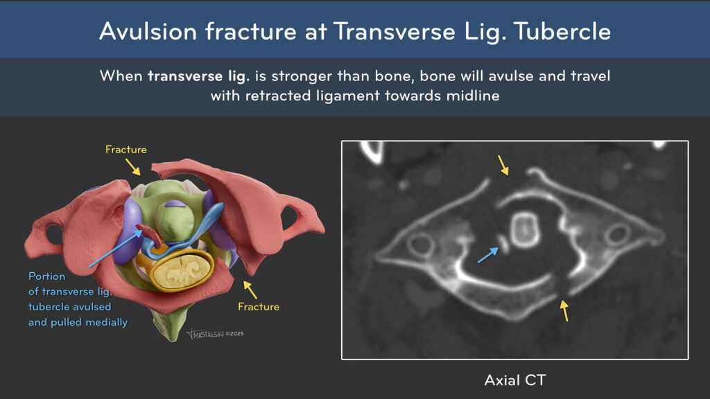
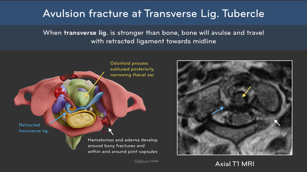
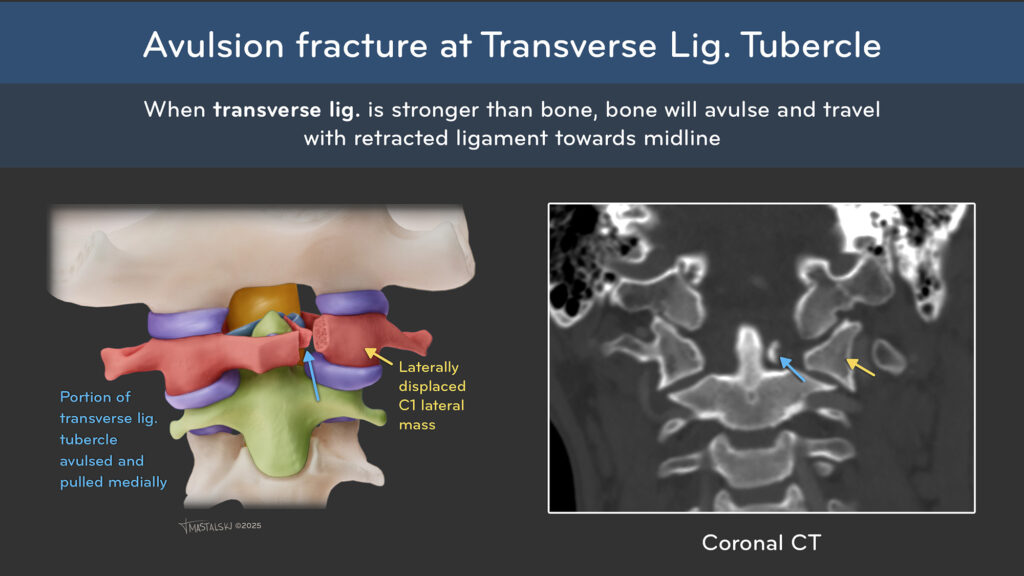
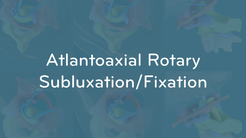
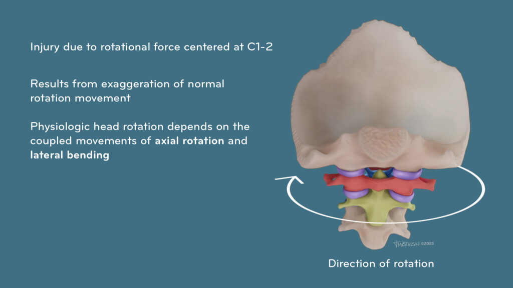
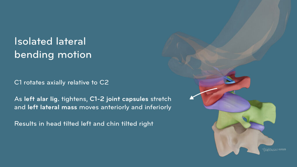
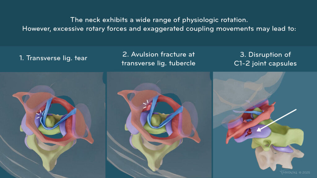
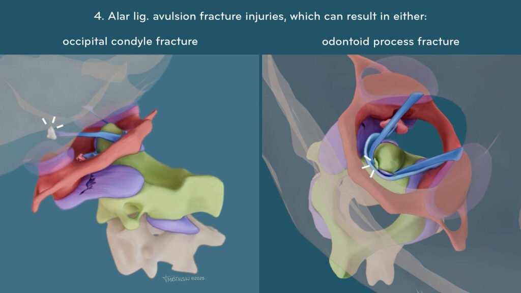
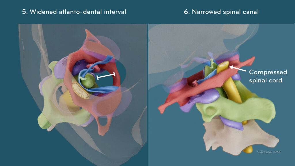
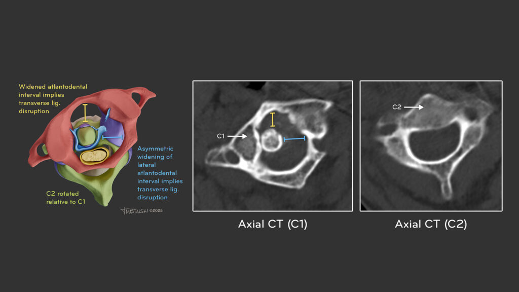
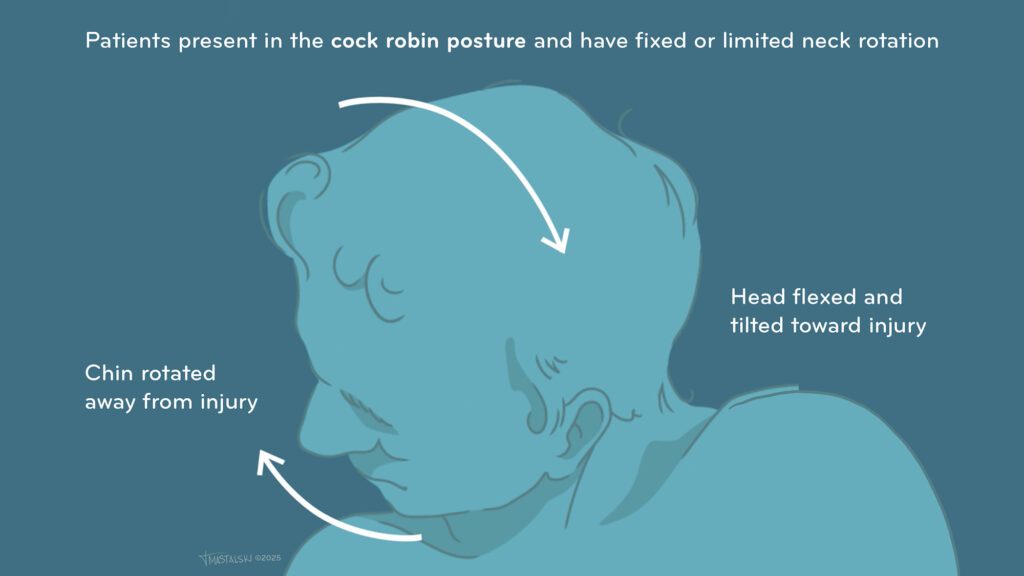
Atlantoaxial Rotary Subluxation/Fixation


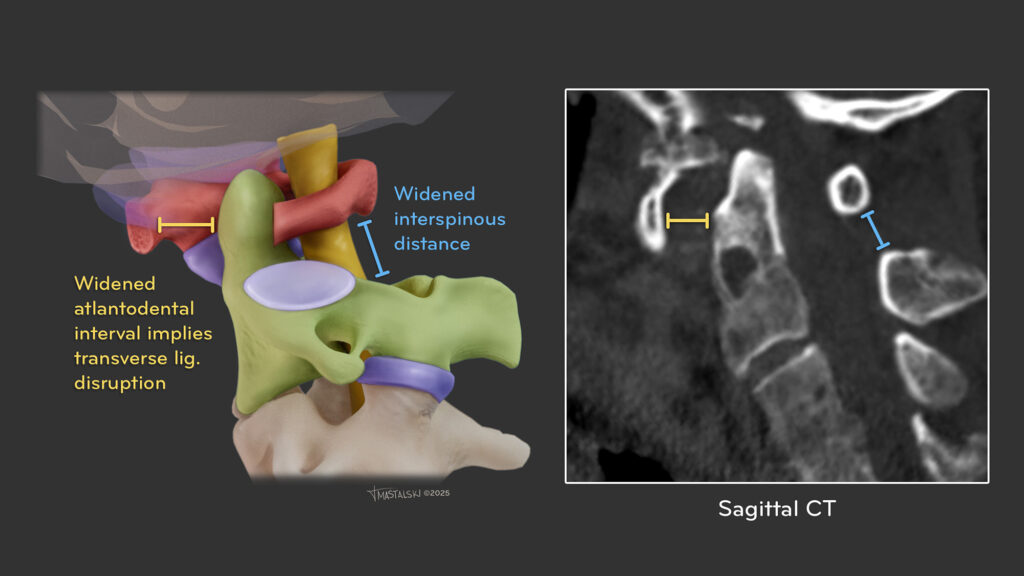
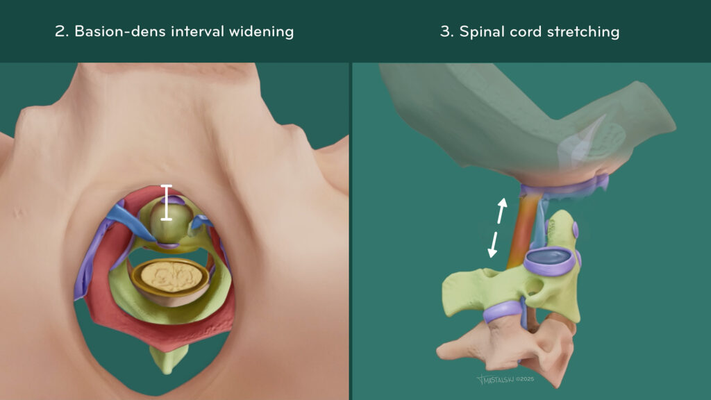
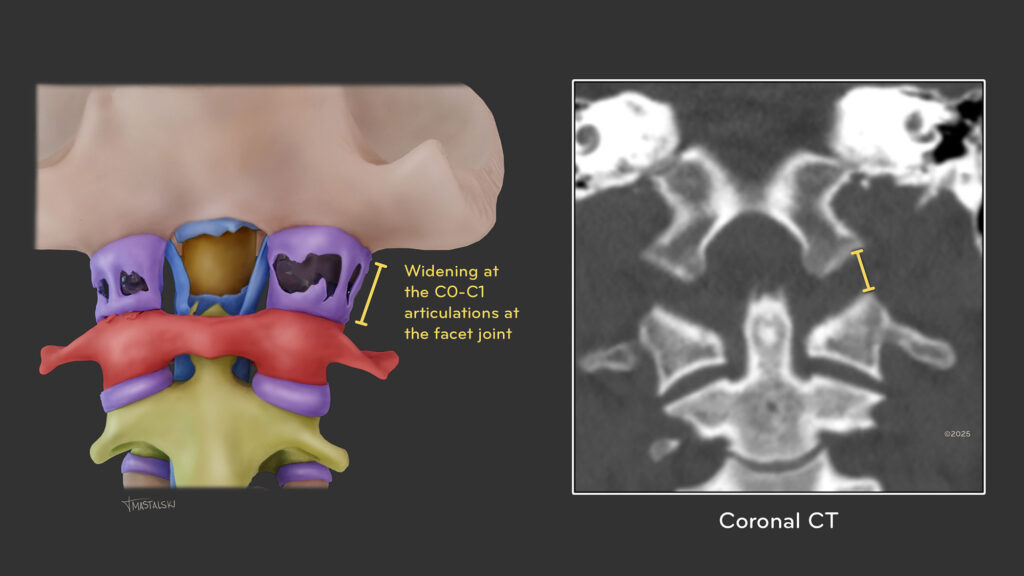
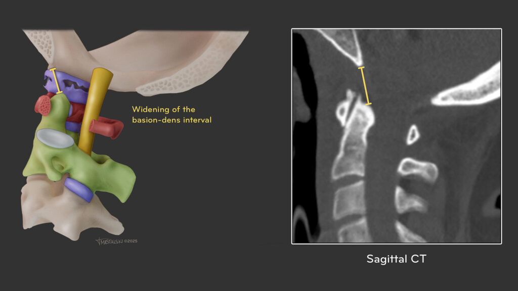
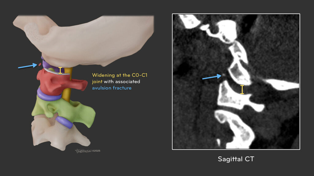
Atlanto-occipital Dissociation